© Copyright Goldendoodles.com 2001. All rights reserved. You may not copy or otherwise use anything on this site without our written permission.

Canine Hip Dysplasia Part I
To understand this genetically transmitted disease, we must first understand the workings of the normal canine hip. By John C. Cargill, MA MBA, MS and Susan Thorpe-Vargas, MS
This is the first in a series of articles addressing canine hip dysplasia. What
follows is written from the perspective that the readers of the series are
conscientious breeders who are the guardians of the genetic pools that constitute
their breeds. While this series of articles will not replace a stack of veterinary
medical texts, it is a relatively in-depth look at the whole problem of canine hip dysplasia.
Furthermore, the series is designed to be retained as a reference. When you finish reading it you
will have a sufficient background to make rational breeding choices and will be able to discuss the
subject from an informed basis with your veterinarian. You may not like what you read, but you will
be more competent to deal with the problem.
Hip dysplasia is one of the most controversial and widespread problems in the dog fancy. So many
old-wives tales, anecdotes, misconceptions and even lies abound that one of the goals of this series
of articles must be to lay things out to the reader as they are, supported with some scientific basis.
Let's start with a hypothetical scenario, but one which too many of us have faced:
He's major-pointed; he moves like a dream; that head piece may just be the best you have ever
bred. In short, this boy typifies everything that is good about your breed and is the culmination of
many years of hard work, hopes, tears, frustration and all the ups and downs, joys and heartaches
common to the fancy. Now it is time to X-ray his hips so that you can not only use him in your
breeding program, but advertise him at stud. This is one boy that is going to make it, and we are
talking national specialty here.
Problem - the radiographic results come back with a diagnosis of canine hip dysplasia-severe. What
should you do?
More among us than will admit have had this experience, and most of those who haven't have seen
it happen to other breeders concentrating on similar bloodlines. Now back to our hypothetical
scenario:
You never suspected a thing. The dog never appeared to be in pain and his gait was what won him
his major points. You have invested time, money and your hopes on this animal, and it all has been
for naught! Now is the time for hysteria and self-blame:
•
What went wrong?
•
Could this have been prevented?
•
Was he not fed correctly?
•
Was he kept on an improper surface while growing?
•
What is this disease that keeps reappearing in the most conscientious of breeding programs,
and which frustrates our attempts to eradicate it?
The first step in understanding canine hip dysplasia is to recognize it as not just one disease but
many diseases, which together result in degenerative effects on the hip joint. An extremely complex
disorder, hip dysplasia is now thought by some to be the most noticeable manifestation of a
systemic condition that can affect not only the hip joints but also those of the elbow, shoulder and
event the joints between the vertebrae1. Whatever else might result from the systemic conditions
of this polygenic and multifactorial disease, hip dysplasia remains a common, usually painful and
often debilitating disease. "Efforts by dog breeders and veterinarians to reduce the prevalence of
the disorder have proven marginally effective." 2
While there is much that we do not know we do know that canine hip dysplasia is a genetically
transmitted disease. If you need to, or if you disagree at this point, please re-read that statement.
We will be repeating it throughout this series of articles, and this concept is the basis for
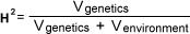
determination of fitness for breeding. The genetic concept of heritability is a complicating factor
and is one reason why hip dysplasia remains so controversial. So often when you breed you get
more than you see. Without resorting to too much math, heritability is equal to the statistical
variance due to genetic influence divided by the sum of the statistical variance due to the genetic
influence plus the variance due to the environmental influence. It is easier to comprehend the
mathematical notation than the statement of the equation:
H2= heritability index
Vgenetics = variance due to genetics
Venvironment = variance due to environmental influences
Thus, heritability is defined as an estimate of how much environmental factors play in the
expression of the inherited genes. A high heritability index means that environmental
considerations are not as important as genetic elements. The numerical value or heritability index is
a function not only of breed type but of the population from which the data is extracted. "Studies of
hip dysplasia genetics have indicated that the disease is polygenic and multifactorial, with estimates
of heritability index in the range of 0.2 to 0.3"3
For instance, in a 1986 Swedish study, the heritability of hip dysplasia in German Shepherds was
0.40 in Sweden, but only 0.25 in the British Isles during the same time period. The difference
between breeds may also reflect their levels of inbreeding. The more inbreeding, the lower the
heritability index because inbreeding reduces the total genetic variability-that is, the gene pool is
smaller. Inbreeding is not a bad word. It only becomes problematic when undesirable genetic traits
are concentrated within the gene pool. By definition, every purebred dog of any given breed is
highly inbred, or else it would look like a feral dog. We frequently hear that the problem with the
American Kennel Club purebred dogs is that they are inbred. We should hope so, otherwise we
could never fix type to the point where there were discernible differences between breeds. On the
other hand, we would hope that the concentrated gene pools for the various breeds would have
been concentrated from stock exhibiting only desirable genetic traits. We would hope that our field,
bench and obedience champions would be fit to contribute to the gene pool. Of course, we know
that is not true, or there would be no purpose in writing this article. 4,5,6
(diagram based on reprint from the Journal of the American Veterinary Association, Vol.196,
No.1,pp.59-70. "New concepts of coxofemoral joint stability and the development of a clinical stress-
radiographic method for quantitating hip joint laxity in the dog," by Gail K. Smith, V.M.D., Ph.D.;
Darryl N. Biery, D.V.M.; and Thomas P. Gregor, B.S.)
To further complicate matters is the fact that the pattern of inheritance indicates that more than one
gene is involved. Hip dysplasia is polygenic (involves many different genes) and multifactorial (influenced
by many non-genetic factors). This makes sense when you think of the complexity of the various structures
involved. Every cell in the body, except for sex cells, carries two copies of each gene and each gene codes
for a specific characteristic. One very simple example is eye color:
If the cell's two sets of genes for a specific characteristic are exactly alike, then the animal is homozygous
for that characteristic.
If the two genes are different, i.e., heterozygous, then one copy of the genes could code for blue eyes and
the other could code for brown eyes.
Let's complicate the matter even further. If the animal carries two different copies of the same gene for
eye color, only one copy can be expressed in any given eye. Closer to home, in humans for example, a
child born to parents heterozygous for eye color (both parents have a blue-eyed gene and brown-eyed
gene) will have a one-in-four chance of having blue eyes. This is because the gene for blue eyes is recessive
and both copies for that code for blue eyes must be present before that characteristic can be expressed.
On the other hand, if the child has brown eyes, we don't know what type of genes for eye color he or she
has. This is because the gene for brown eyes is dominant and is able to "mask" the physical expression of
the blue-eyed gene. Alternatively, the child could have only the genes that code for brown eyes. It is
beyond the scope of this article to address the various "odd" eye color combinations, but co-dominance
and variable penetrance may be what we are dealing with in canine hip dysplasia.
What you have just read is an example of phenotype vs. Genotype. Phenotype is the physical expression
of a genetic characteristic. Genotype is genetic composition of the organism. Using our eye-color
example, the child with two different copies of the gene will express the brown-eyed phenotype, but
his or her genotype will be heterozygous.
Let's add to the complexity once again. Co-dominance of genes is a situation where neither gene is
dominant. A clear example illustrating the concept of genetic co-dominance is flower color. A snap
dragon homozygous (both copies of color genes exactly alike) for white petals crossed with a snap
dragon homozygous for red petals will produce a flower with pink petals, not a flower with either
white or red petals or a mixture of red and white petals. Many researchers feel that hip dysplasia
may be a mixture of dominant, recessive and co-dominant genes. Quite probably, this is one of the
reasons why isolation of the causative genetic factors of canine hip dysplasia has been so elusive.
The concepts that you need to be clear on as we leave this mini-course on genetics are: heritability
index; genetic and environmental variability; dominant vs. Recessive genes; homozygous vs.
heterozygous; genetic co-dominance; and most importantly that hip dysplasia is genetically
inheritable and is polygenic and multifactorial. In short, you can get it in your breeding program
when you bred from animals that did not show it.
Before we can discuss an abnormal process (disease), we need to first understand the normal
process. In this case, we must be able to answer the question, "What is a normal hip, what makes it
normal, and how does it get that way?"
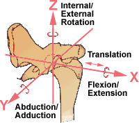
First, what is the hip? The hip joint is a main weight-bearing joint consisting principally of a ball and
socket. This joint connects the pelvis to the lower extremities. The ball is on the end of the femur
(thigh bone) and the socket (acetabulum) is part of the pelvis. Note from figure 1 how the femoral
head fits into the acetabulum in the normal hip joint. This will be key to all our discussions from this
point forth. A true ball-and-socket joint has three degrees of freedom, that is, it supports rotation
about three different axes. The canine hip joint is unusual as a ball-and-socket joint in that it has a
fourth degree of freedom. The femoral head may be displaced laterally from the acetabulum. While
this is the genius of this joint, allowing the attached appendage a full range of motion, it can also
create a problem if there becomes too much laxity in the joint. Note the fourth degree of freedom
in Figure 2, which provides for the femoral head (ball) to move directly away from the acetabulum
(socket). From Figures 1 and 2, it should be obvious that much lateral displacement of the femoral
head from its seat in the acetabulum will produce high joint stresses during weight bearing. This
joint laxity will be a major consideration for the changes it causes in the joint mechanics as we
progress through this series of articles.
The acetabulum is formed from the embryonic process of fusion of the ilium (top of the hip), the
ischium (lowest part of the hip) and the pubis (below the ilium but above the pubis) and the
acetabular bone. Most researchers feel that normal development requires close conformity (close,
tight fit) between the acetabulum and the femoral head throughout their growth period. In other
words, the joint must fit tightly, deeply and snugly. This is how a puppy's hip starts out-dysplastic
and non-dysplastic puppies' hips are indistinguishable. The first six months of life seem to be the
most critical growth period when the depth of the socket must be maintained. It is believed that the
depth of the socket in the growing puppy may be in part a function of the amount of stress the
femoral head can produce on the immature acetabulum. Think of it as a thumb pushing into a ball
of clay. The harder the thumb pushes, the deeper the indentation in the clay. Much as a knife edge
concentrates force onto a relatively small surafce area (and a pin of a diameter equal to the width of
the knife edge even more), the two phenotypic traits that maximize the forces between these two
developing bony structures are a small femoral head and a long femoral neck. Note, however, that
the normal acetabulum is well-formed in utero, thus the stress may only serve to maintain that
socket depth.
To cushion the force between these two bony surfaces, there is a truly remarkable substance called
articular cartilage. This cartilage is similar to a hard sponge with a slick hard surface facing the
interior of the joint. In the normal joint, articular cartilage is able to change its shape slightly when
force is applied to it, thus spreading and distributing force more evenly into the subchrondal bone
directly beneath the articular cartilage. This is of major importance to the long-term integrity of the
joint.
Holding everything in place is another structure that does more than just enhance the stability of
the joint. The joint capsule is a fibrous structure filled with synovial fluid that surrounds, isolates
and protects the joint. This joint capsule is essential to proper development and functioning of the
joint. This structure is similar to the rubber grease bladder around a ball joint in the front
suspension of your car. The cushioning effect of the grease with the fluid pressure of the grease
and the elasticity of the bladder helps to stabilize the joint. The bladder helps keep out
contaminants. This function becomes even more important as the joint ages and surfaces become
worn. The joint capsule contains the all-important synovial fluid, the most important ingredients of
which are nutrients, which diffuse into the joint from the blood supply, and hyaluronic acid (HA).
The tissues within the joint extract nutrients from the synovial fluid in which they are bathed.
Hyaluronic acid has a critical function: to provide lubrication. This slippery and viscous substance
prevents rapid erosion of the articular cartilage and the surfaces of the femoral head and the
acetabulum. A membrane called the synovial membrane lines the inside of the joint capsule,
providing further isolation of the joint space. Should the synovial membrane become injured or
ruptured, white blood cells release enzymes and oxygen radials (free radicals) that attack and
destroy hyaluronic acid. When this occurs, the loss of HA reduces the lubrication that prevents
friction and limits erosion of the articular cartilage. Even worse, loss of HA allows the enzymes from
white blood cells to join forces with oxygen free radicals and attack the articular cartilage. Free
radicals play a major role in degenerative arthritis.
The ball-and-socket (coxofemoral) joints of an affected puppy radiographically appear to be
structurally and functionally normal at birth. The hips of an affected puppy are indistinguishable
from a normal puppy at birth. This is an important point to remember. As an affected puppy grows,
the hip joint undergoes severe structural alterations. The changes result from joint laxity and
adulteration/destruction of the constituents of the synovial fluid and subsequent loss of lubrication
and nourishment, which serve to reduce the regenerative and elastic (force-absorbing and
distributing) properties of the articular cartilage. The normal joint retains its tightness and close fit.
Whereas in the genetically dysplastic-to-be puppy, the acetabular rim and femoral head become
eroded.
Remember that the acetabular depth is partially a function of the small "footprint" of the femoral
head which concentrates force into a small surface area. As the femoral head is flattened, the
coxofemoral joint no longer fits snugly. Excessive force is applied unevenly, especially at the edges
of the flattened femoral head. Visualize this joint looseness as the difference between the impact of
a boxer's fist when the punch is thrown with the glove already in contact with the opponent's jaw as
contrasted with an initial stand-off distance of say 20 inches. In the first case, little impact force is
transmitted and no damage is done; in the second, there may be a knock-out. In the joint, the
increase in stress results not only in abnormal wear of the articular cartilage, but causes tiny micro-
stress fractures to appear in the subchondral bone. The body attempts to heal these fissures,
causing the acetabulum to become filled in, i.e., made shallower. It is this cycle of damage and
repair (osteophyte formation) that leads to deformation of the joint, and degenerative hip disease.
Conclusions: Hip dysplasia is not something a dog acquires; a dog either is genetically dysplastic or
it is not. Initially, the hips of affected and normal puppies are indistinguishable. Later in life, an
affected animal can exhibit a wide range of phenotypes, all the way from normal to severely
dysplastic and functionally crippled. You should take away from this article the idea that hip
dysplasia is genetically inherited. Never believe a fellow breeder or fancier who claims there is no
hip dysplasia in his or her line. Never believe breeders who claim that if their breeding lines carried
the genes for hip dysplasia they would be able to see it in their animals' gaits. This just is not true.
Although work has been started to find the genetic markers for the disease, we have as yet no
method of genetic analysis that can tell breeders whether their dogs are dysplastic or not. We only
have physical expression of the disease, and an effort to "back door" into clear stock for breeding
purposes. Breeders must come to understand that the only way to reduce the incidence of hip
dysplasia is by trying to breed from as few animals that have progenitors, siblings, get, or get of
siblings that had clinical manifestations of hip dysplasia. Obviously, a great amount of information
is lacking to make a rational breeding choice. These are hard words to have read, but much of our
problem has come from thousands of years of less than natural selection resulting from the
domestication of the dog.
In our second article in this series we will address in greater detail the parts nutritional,
environmental and other factors play in mitigating or increasing the physical expression of canine
hip dysplasia.
CREDITS
References
1. Olsewski J.M., Lust G., Rendano B.T., et al. "Degenerative joint disease: Multiple joint involvement
in young and mature dogs." Am J Vet Res. 1983; vol 44:1300-1308.
2. Smith G.K., Biery D.N. "New concepts of coxofemoral joint stability and the development of a
clinical stress-radiographic method for quantitating hip joint laxity in the dog." J Am Vet Med Assoc.
1990;196:59-70.
3. Ibid., p. 59.
4. Cargill J. "Truth in advertising: breeder self-regulation I." Dog World. 1990(Jul);75 No.7:38-82.
5. Cargill J. "Truth in advertising: breeder self-regulation II." Dog World. 1990(Aug);75 No.8:111-116.
6. Cargill J. "What should 'champion' mean?" Dog World. 1993(Feb);78 No.2:34-35.
The information contained on this site is in no way intended to replace that of proper veterinary advice, diagnosis or treatment.
It is meant to provide resource, so that we can better understand canine health related issues.

© Copyright Goldendoodles.com 2001. All rights reserved. You may not
copy or otherwise use anything on this site without our written permission
Made with Xara


The information contained on this site is in no way intended to
replace that of proper veterinary advice, diagnosis or treatment.
It is meant to provide resource, so that we can better understand
canine health related issues.
Canine Hip Dysplasia Part I
To understand this genetically transmitted disease, we must first understand the workings of the normal canine hip. By John C. Cargill, MA MBA, MS and Susan Thorpe-Vargas, MS
This is the first in a series of articles addressing canine hip
dysplasia. What follows is written from the perspective that the
readers of the series are conscientious breeders who are the
guardians of the genetic pools that constitute their breeds. While
this series of articles will not replace a stack of veterinary
medical texts, it is a relatively in-depth look at the whole
problem of canine hip dysplasia. Furthermore, the series is
designed to be retained as a reference. When you finish reading
it you will have a sufficient background to make rational
breeding choices and will be able to discuss the subject from an
informed basis with your veterinarian. You may not like what
you read, but you will be more competent to deal with the
problem.
Hip dysplasia is one of the most controversial and widespread
problems in the dog fancy. So many old-wives tales, anecdotes,
misconceptions and even lies abound that one of the goals of
this series of articles must be to lay things out to the reader as
they are, supported with some scientific basis.
Let's start with a hypothetical scenario, but one which too many
of us have faced:
He's major-pointed; he moves like a dream; that head piece may
just be the best you have ever bred. In short, this boy typifies
everything that is good about your breed and is the culmination
of many years of hard work, hopes, tears, frustration and all the
ups and downs, joys and heartaches common to the fancy. Now
it is time to X-ray his hips so that you can not only use him in
your breeding program, but advertise him at stud. This is one
boy that is going to make it, and we are talking national specialty
here.
Problem - the radiographic results come back with a diagnosis of
canine hip dysplasia-severe. What should you do?
More among us than will admit have had this experience, and
most of those who haven't have seen it happen to other
breeders concentrating on similar bloodlines. Now back to our
hypothetical scenario:
You never suspected a thing. The dog never appeared to be in
pain and his gait was what won him his major points. You have
invested time, money and your hopes on this animal, and it all
has been for naught! Now is the time for hysteria and self-
blame:
•
What went wrong?
•
Could this have been prevented?
•
Was he not fed correctly?
•
Was he kept on an improper surface while growing?
•
What is this disease that keeps reappearing in the most
conscientious of breeding programs, and which frustrates
our attempts to eradicate it?
The first step in understanding canine hip dysplasia is to
recognize it as not just one disease but many diseases, which
together result in degenerative effects on the hip joint. An
extremely complex disorder, hip dysplasia is now thought by
some to be the most noticeable manifestation of a systemic
condition that can affect not only the hip joints but also those of
the elbow, shoulder and event the joints between the
vertebrae1. Whatever else might result from the systemic
conditions of this polygenic and multifactorial disease, hip
dysplasia remains a common, usually painful and often
debilitating disease. "Efforts by dog breeders and veterinarians
to reduce the prevalence of the disorder have proven marginally
effective." 2
While there is much that we do not know we do know that
canine hip dysplasia is a genetically transmitted disease. If you
need to, or if you disagree at this point, please re-read that
statement. We will be repeating it throughout this series of
articles, and this concept is the basis for determination of fitness
for breeding. The genetic concept of heritability is a complicating
factor and is one reason why hip dysplasia remains so
controversial. So often when you breed you get more than you
see. Without resorting to too much math, heritability is equal to
the statistical variance due to genetic influence divided by the
sum of the statistical variance due to the genetic influence plus
the variance due to the environmental influence. It is easier to
comprehend the mathematical notation than the statement of
the equation:
H2= heritability index
Vgenetics = variance due to genetics
Venvironment = variance due to environmental influences
Thus, heritability is defined as an estimate of how much
environmental factors play in the expression of the inherited
genes. A high heritability index means that environmental
considerations are not as important as genetic elements. The
numerical value or heritability index is a function not only of
breed type but of the population from which the data is
extracted. "Studies of hip dysplasia genetics have indicated that
the disease is polygenic and multifactorial, with estimates of
heritability index in the range of 0.2 to 0.3"3
For instance, in a 1986 Swedish study, the heritability of hip
dysplasia in German Shepherds was 0.40 in Sweden, but only
0.25 in the British Isles during the same time period. The
difference between breeds may also reflect their levels of
inbreeding. The more inbreeding, the lower the heritability index
because inbreeding reduces the total genetic variability-that is,
the gene pool is smaller. Inbreeding is not a bad word. It only
becomes problematic when undesirable genetic traits are
concentrated within the gene pool. By definition, every purebred
dog of any given breed is highly inbred, or else it would look like
a feral dog. We frequently hear that the problem with the
American Kennel Club purebred dogs is that they are inbred. We
should hope so, otherwise we could never fix type to the point
where there were discernible differences between breeds. On
the other hand, we would hope that the concentrated gene
pools for the various breeds would have been concentrated
from stock exhibiting only desirable genetic traits. We would
hope that our field, bench and obedience champions would be
fit to contribute to the gene pool. Of course, we know that is not
true, or there would be no purpose in writing this article. 4,5,6
(diagram based on reprint from the Journal of the American
Veterinary Association, Vol.196, No.1,pp.59-70. "New concepts of
coxofemoral joint stability and the development of a clinical
stress-radiographic method for quantitating hip joint laxity in the
dog," by Gail K. Smith, V.M.D., Ph.D.; Darryl N. Biery, D.V.M.; and
Thomas P. Gregor, B.S.)
To further complicate matters is the fact that the pattern of
inheritance indicates that more than one gene is involved. Hip
dysplasia is polygenic (involves many different genes) and
multifactorial (influenced by many non-genetic factors). This makes
sense when you think of the complexity of the various structures
involved. Every cell in the body, except for sex cells, carries two
copies of each gene and each gene codes for a specific
characteristic. One very simple example is eye color:
If the cell's two sets of genes for a specific characteristic are exactly
alike, then the animal is homozygous for that characteristic.
If the two genes are different, i.e., heterozygous, then one copy of the
genes could code for blue eyes and the other could code for brown
eyes.
Let's complicate the matter even further. If the animal carries two
different copies of the same gene for eye color, only one copy can be
expressed in any given eye. Closer to home, in humans for example,
a child born to parents heterozygous for eye color (both parents
have a blue-eyed gene and brown-eyed gene) will have a one-in-four
chance of having blue eyes. This is because the gene for blue eyes is
recessive and both copies for that code for blue eyes must be
present before that characteristic can be expressed. On the other
hand, if the child has brown eyes, we don't know what type of genes
for eye color he or she has. This is because the gene for brown eyes
is dominant and is able to "mask" the physical expression of the
blue-eyed gene. Alternatively, the child could have only the genes
that code for brown eyes. It is beyond the scope of this article to
address the various "odd" eye color combinations, but co-
dominance and variable penetrance may be what we are dealing
with in canine hip dysplasia.
What you have just read is an example of phenotype vs. Genotype.
Phenotype is the physical expression of a genetic characteristic.
Genotype is genetic composition of the organism. Using our eye-
color example, the child with two different copies of the gene will
express the brown-eyed phenotype, but his or her genotype will
be heterozygous.
Let's add to the complexity once again. Co-dominance of genes
is a situation where neither gene is dominant. A clear example
illustrating the concept of genetic co-dominance is flower color.
A snap dragon homozygous (both copies of color genes exactly
alike) for white petals crossed with a snap dragon homozygous
for red petals will produce a flower with pink petals, not a flower
with either white or red petals or a mixture of red and white
petals. Many researchers feel that hip dysplasia may be a
mixture of dominant, recessive and co-dominant genes. Quite
probably, this is one of the reasons why isolation of the
causative genetic factors of canine hip dysplasia has been so
elusive.
The concepts that you need to be clear on as we leave this mini-
course on genetics are: heritability index; genetic and
environmental variability; dominant vs. Recessive genes;
homozygous vs. heterozygous; genetic co-dominance; and most
importantly that hip dysplasia is genetically inheritable and is
polygenic and multifactorial. In short, you can get it in your
breeding program when you bred from animals that did not
show it.
Before we can discuss an abnormal process (disease), we need
to first understand the normal process. In this case, we must be
able to answer the question, "What is a normal hip, what makes
it normal, and how does it get that way?"
First, what is the hip? The hip joint is a main weight-bearing joint
consisting principally of a ball and socket. This joint connects the
pelvis to the lower extremities. The ball is on the end of the
femur (thigh bone) and the socket (acetabulum) is part of the
pelvis. Note from figure 1 how the femoral head fits into the
acetabulum in the normal hip joint. This will be key to all our
discussions from this point forth. A true ball-and-socket joint has
three degrees of freedom, that is, it supports rotation about
three different axes. The canine hip joint is unusual as a ball-
and-socket joint in that it has a fourth degree of freedom. The
femoral head may be displaced laterally from the acetabulum.
While this is the genius of this joint, allowing the attached
appendage a full range of motion, it can also create a problem if
there becomes too much laxity in the joint. Note the fourth
degree of freedom in Figure 2, which provides for the femoral
head (ball) to move directly away from the acetabulum (socket).
From Figures 1 and 2, it should be obvious that much lateral
displacement of the femoral head from its seat in the
acetabulum will produce high joint stresses during weight
bearing. This joint laxity will be a major consideration for the
changes it causes in the joint mechanics as we progress through
this series of articles.
The acetabulum is formed from the embryonic process of fusion
of the ilium (top of the hip), the ischium (lowest part of the hip)
and the pubis (below the ilium but above the pubis) and the
acetabular bone. Most researchers feel that normal
development requires close conformity (close, tight fit) between
the acetabulum and the femoral head throughout their growth
period. In other words, the joint must fit tightly, deeply and
snugly. This is how a puppy's hip starts out-dysplastic and non-
dysplastic puppies' hips are indistinguishable. The first six
months of life seem to be the most critical growth period when
the depth of the socket must be maintained. It is believed that
the depth of the socket in the growing puppy may be in part a
function of the amount of stress the femoral head can produce
on the immature acetabulum. Think of it as a thumb pushing
into a ball of clay. The harder the thumb pushes, the deeper the
indentation in the clay. Much as a knife edge concentrates force
onto a relatively small surafce area (and a pin of a diameter
equal to the width of the knife edge even more), the two
phenotypic traits that maximize the forces between these two
developing bony structures are a small femoral head and a long
femoral neck. Note, however, that the normal acetabulum is
well-formed in utero, thus the stress may only serve to maintain
that socket depth.
To cushion the force between these two bony surfaces, there is a
truly remarkable substance called articular cartilage. This
cartilage is similar to a hard sponge with a slick hard surface
facing the interior of the joint. In the normal joint, articular
cartilage is able to change its shape slightly when force is applied
to it, thus spreading and distributing force more evenly into the
subchrondal bone directly beneath the articular cartilage. This is
of major importance to the long-term integrity of the joint.
Holding everything in place is another structure that does more
than just enhance the stability of the joint. The joint capsule is a
fibrous structure filled with synovial fluid that surrounds, isolates
and protects the joint. This joint capsule is essential to proper
development and functioning of the joint. This structure is
similar to the rubber grease bladder around a ball joint in the
front suspension of your car. The cushioning effect of the grease
with the fluid pressure of the grease and the elasticity of the
bladder helps to stabilize the joint. The bladder helps keep out
contaminants. This function becomes even more important as
the joint ages and surfaces become worn. The joint capsule
contains the all-important synovial fluid, the most important
ingredients of which are nutrients, which diffuse into the joint
from the blood supply, and hyaluronic acid (HA). The tissues
within the joint extract nutrients from the synovial fluid in which
they are bathed.
Hyaluronic acid has a critical function: to provide lubrication.
This slippery and viscous substance prevents rapid erosion of
the articular cartilage and the surfaces of the femoral head and
the acetabulum. A membrane called the synovial membrane
lines the inside of the joint capsule, providing further isolation of
the joint space. Should the synovial membrane become injured
or ruptured, white blood cells release enzymes and oxygen
radials (free radicals) that attack and destroy hyaluronic acid.
When this occurs, the loss of HA reduces the lubrication that
prevents friction and limits erosion of the articular cartilage.
Even worse, loss of HA allows the enzymes from white blood
cells to join forces with oxygen free radicals and attack the
articular cartilage. Free radicals play a major role in degenerative
arthritis.
The ball-and-socket (coxofemoral) joints of an affected puppy
radiographically appear to be structurally and functionally
normal at birth. The hips of an affected puppy are
indistinguishable from a normal puppy at birth. This is an
important point to remember. As an affected puppy grows, the
hip joint undergoes severe structural alterations. The changes
result from joint laxity and adulteration/destruction of the
constituents of the synovial fluid and subsequent loss of
lubrication and nourishment, which serve to reduce the
regenerative and elastic (force-absorbing and distributing)
properties of the articular cartilage. The normal joint retains its
tightness and close fit. Whereas in the genetically dysplastic-to-
be puppy, the acetabular rim and femoral head become eroded.
Remember that the acetabular depth is partially a function of
the small "footprint" of the femoral head which concentrates
force into a small surface area. As the femoral head is flattened,
the coxofemoral joint no longer fits snugly. Excessive force is
applied unevenly, especially at the edges of the flattened
femoral head. Visualize this joint looseness as the difference
between the impact of a boxer's fist when the punch is thrown
with the glove already in contact with the opponent's jaw as
contrasted with an initial stand-off distance of say 20 inches. In
the first case, little impact force is transmitted and no damage is
done; in the second, there may be a knock-out. In the joint, the
increase in stress results not only in abnormal wear of the
articular cartilage, but causes tiny micro-stress fractures to
appear in the subchondral bone. The body attempts to heal
these fissures, causing the acetabulum to become filled in, i.e.,
made shallower. It is this cycle of damage and repair (osteophyte
formation) that leads to deformation of the joint, and
degenerative hip disease.
Conclusions: Hip dysplasia is not something a dog acquires; a
dog either is genetically dysplastic or it is not. Initially, the hips of
affected and normal puppies are indistinguishable. Later in life,
an affected animal can exhibit a wide range of phenotypes, all
the way from normal to severely dysplastic and functionally
crippled. You should take away from this article the idea that hip
dysplasia is genetically inherited. Never believe a fellow breeder
or fancier who claims there is no hip dysplasia in his or her line.
Never believe breeders who claim that if their breeding lines
carried the genes for hip dysplasia they would be able to see it in
their animals' gaits. This just is not true.
Although work has been started to find the genetic markers for
the disease, we have as yet no method of genetic analysis that
can tell breeders whether their dogs are dysplastic or not. We
only have physical expression of the disease, and an effort to
"back door" into clear stock for breeding purposes. Breeders
must come to understand that the only way to reduce the
incidence of hip dysplasia is by trying to breed from as few
animals that have progenitors, siblings, get, or get of siblings
that had clinical manifestations of hip dysplasia. Obviously, a
great amount of information is lacking to make a rational
breeding choice. These are hard words to have read, but much
of our problem has come from thousands of years of less than
natural selection resulting from the domestication of the dog.
In our second article in this series we will address in greater
detail the parts nutritional, environmental and other factors play
in mitigating or increasing the physical expression of canine hip
dysplasia.
CREDITS
References
1. Olsewski J.M., Lust G., Rendano B.T., et al. "Degenerative joint
disease: Multiple joint involvement in young and mature dogs."
Am J Vet Res. 1983; vol 44:1300-1308.
2. Smith G.K., Biery D.N. "New concepts of coxofemoral joint
stability and the development of a clinical stress-radiographic
method for quantitating hip joint laxity in the dog." J Am Vet Med
Assoc. 1990;196:59-70.
3. Ibid., p. 59.
4. Cargill J. "Truth in advertising: breeder self-regulation I." Dog
World. 1990(Jul);75 No.7:38-82.
5. Cargill J. "Truth in advertising: breeder self-regulation II." Dog
World. 1990(Aug);75 No.8:111-116.
6. Cargill J. "What should 'champion' mean?" Dog World.
1993(Feb);78 No.2:34-35.